Figure 1.

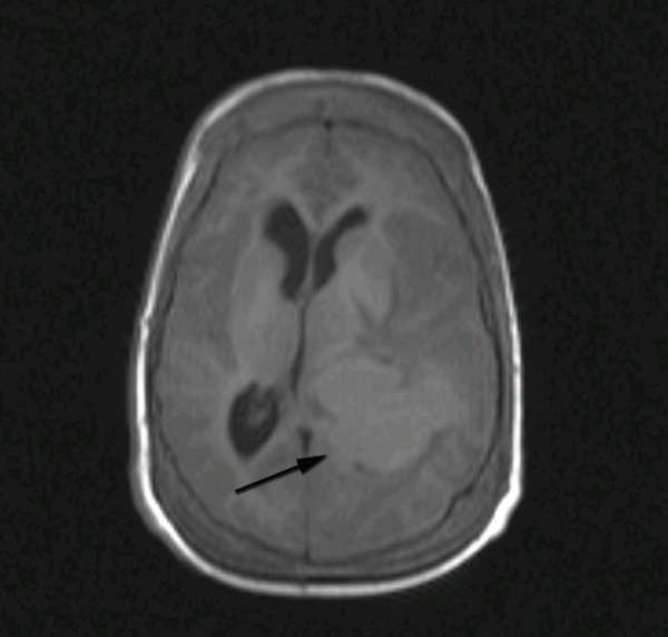
Axial T1-weighted MRI image demonstrating a well-demarcated isointense lesion in the left trigone with significant surrounding edema and mass effect.

Axial T1-weighted MRI image demonstrating a well-demarcated isointense lesion in the left trigone with significant surrounding edema and mass effect.