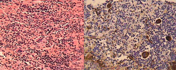
Figure 4.

Microscopic view of the excised brain lesion. Demonstrates (A) the infiltration by foci of hematopoietic cells and megakaryocytes (hematoxylin and eosin stain, original magnification ×40) and (B) megakaryocytes and endothelial cells positive for factor VIII related antigen (hematoxylin and eosin stain, original magnification ×40).
