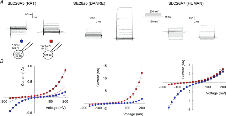
Figure 3. SCN− currents in cells stably expressing mammalian prestin, teleost prestin and human SLC26A7.

A, representative whole-cell SCN− currents in Flp-In T-REx cells stably expressing YFP-tagged SLC26A5 (RAT)/prestin, Slc26a5 (DANRE)/prestin or human SLC26A7 for two different anion distributions shown in the cartoon. The dotted lines indicate the zero current reference level. B, current–voltage relationships for YFP-tagged SLC26A5 (RAT)/prestin, Slc26a5 (DANRE)/prestin or human SLC26A7 determined by current recordings as shown in A. Means ± SEM from more than 7 cells. Protein expression was induced with tetracycline 24 h before electrophysiological characterization for the two prestins and 8 h before characterization of SLC26A7.
