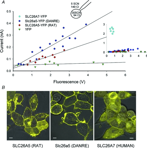
Figure 4. Comparison of SCN− transport by different SLC26s.

A, plot of whole-cell current amplitudes at –110 mV versus whole-cell fluorescence for Flip-In T-REx cells expressing YFP-tagged SLC26A5 (RAT)/prestin, Slc26a5 (DANRE)/prestin or human SLC26A7 (inset). An inducible stable cell line expressing only YFP was used as negative control. B, confocal images of Flp-In T-REx cells expressing the indicated YFP-tagged proteins superimposed on differential interference contrast images. Cells were incubated with tetracycline for either 24–48 h (SLC26A5 (RAT), Slc26a5 (DANRE)) or 8 h (YFP, human SLC26A7). Bar = 5 μm.
