Figure 3.
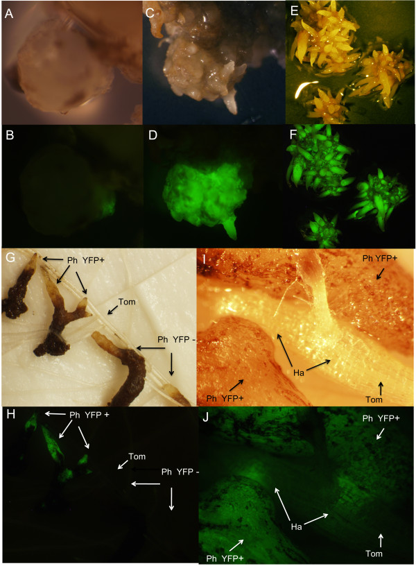
Selection and regeneration of transformed callus. White light (A, C, E, G, and I) and UV illuminated (B, D, F, H, and J) P. aegyptiaca tissues showing YFP fluorescence. A and B) in vitro callus of P. aegyptiaca expressing a small sector of YFP on day 35 after transformation. C and D) chimeric transgenic P. aegyptiaca on day 90 after transformation. YFP-negative tissue was cut away and the YFP- positive tissue subjected to clonal propagation resulting in homogeneus transgenic calli seen in E and F. E and F) clonally propagated calli used for tomato inoculation on day 120 after transformation. G and H) tomato inoculation with excised explants from the transgenic calli. YFP-positive and negative parasitic roots were inoculated next to each other to show the absence of fluorescence in tomato roots and YFP-negative explants. I and J) Detail image of tomato host root parasitized by P. aegyptiaca explants from both sides. The wedge-shaped haustoria penetrating the host root are visible through the intact tomato living tissue. Ph YFP+, P. aegyptiaca positive expressing YFP; Ph YFP-, P. aegyptiaca negative control; Tom, tomato root; Ha, YFP positive haustoria.

