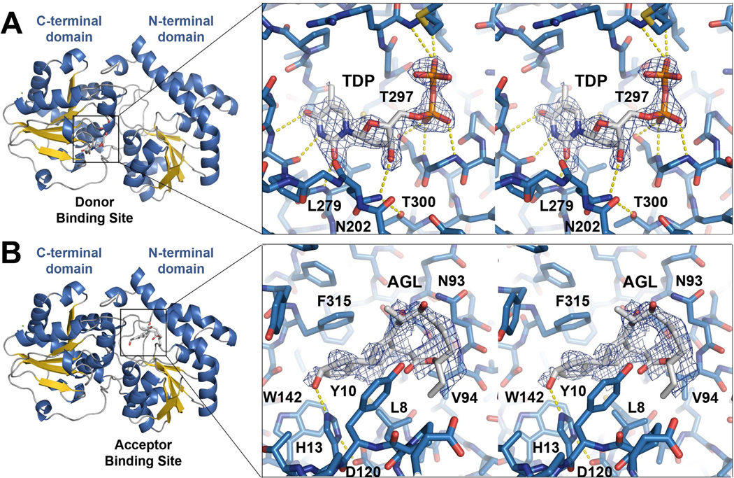
Figure 3.
The donor and acceptor binding sites. a) A stereodiagram of the donor binding site of the SpnG-TDP complex shows the simulated-annealing Fo−Fc omit map of TDP (contoured at 2.5 σ). The thymine base, deoxyribose, and the pyrophosphate of TDP form hydrogen bonds with the donor binding site. b) A stereodiagram of the acceptor binding site of the SpnG-AGL complex shows the simulated-annealing Fo−Fc omit map of AGL (contoured at 2.5 σ). Though AGL is a substrate mimic, the putative natural substrates 3 and 4 likely bind similarly via hydrophobic contacts.

