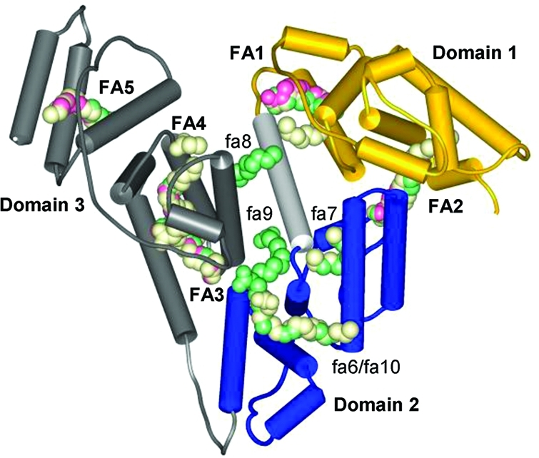
Figure 1.

Domain structure of albumin and fatty acid binding sites. Overlaid structures with PDB codes: 1bj5, HSA with five myristates, pink (the protein backbone is also shown); 1e7e, HSA with 10 decanoates, green; 1gnj, HSA with seven arachidonates, light-yellow.12 FA1–5, major sites; fa6–10, minor sites.
