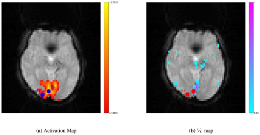
Figure 3. Regions of interest.
Because BOLD contrast is highly weighted by venous blood content, activation areas often overlap with large vein regions. Two regions of interest (ROIs) were selected from visual cortex according to activation detection (warm color) and vascular information (cool color). The spatial resolution of venography map was downsample to identical with that of fMRI image. ( : Primary visual cortex;
  : Visual area
  ).
).

