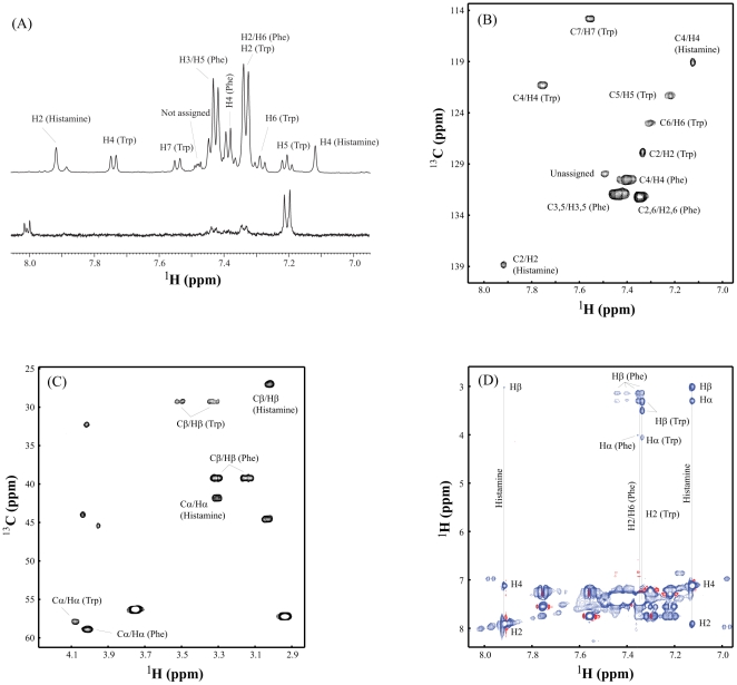
Figure 1. Identification of histamine in TNF-inhibitory fractions of L. reuteri 6475 by NMR.
1H NMR analysis demonstrated differences in the composition of HILIC-HPLC fractions from L. reuteri 6475. A. Top and bottom spectra show the 1D 1H NMR of TNF-inhibitory fraction B3 and fraction B4, respectively. The assigned peaks of phenylalanine (Phe), tryptophan (Trp), and histamine from fraction B3 are labeled on the top spectrum. Two complementary 2D NMR techniques were used to identify compounds unique to TNF-inhibitory fraction B3. B. Aromatic peaks of 1H-13C-HSQC from Phe, Trp, and histamine. C. Aliphatic peaks of 1H-13C-HSQC from Phe, Trp, and histamine. D. The TOCSY spectrum further identified the spin system of Phe, Trp, and histamine unique to TNF-inhibitory fraction B3.

