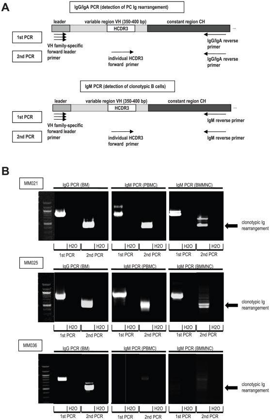
Figure 1. Detection of clonotypic Ig rearrangements by semi-nested PCR.
A: Illustration of semi-nested PCR approach for the detection of clonotypic Ig rearrangements. Ig genes were amplified using VH family-specific forward leader primers and constant region specific reverse primers (1st PCR). A secondary PCR was used to specifically amplify the clonotypic Ig rearrangement with a patient-individual HCDR3-specific primer. PC = plasma cell, Ig = immunoglobulin, VH = heavy chain variable region, CH = heavy chain constant region, HCDR3 = heavy chain complementarity determining region 3. B: Detection of clonotypic Ig-rearrangements in myeloma PBMCs and BMMNCs of patients MM021, MM025 and MM036. Semi-nested PCRs were performed as described in A. PCR products were loaded on agarose gels stained with ethidium bromide. MM = Multiple Myeloma, PBMC = peripheral blood mononuclear cell, BMMNC = bone marrow mononuclear cell.

