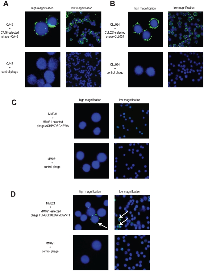
Figure 3. Detection of surface Ig-positive clonotypic B cells by immunofluorescence using patient-individual ligands.
A and B: Ig-selected phage bind to surface Ig-positive parental B cells as visualized by immunofluorescence. As proof of principal an immunofluorescence staining protocol was set up using two different monoclonal B cell systems as positive controls as described in the Methods and Results sections. Phage-CA46 selected on the Ig of the Burkitt lymphoma cell line CA46 (panel A, upper row) as well as control phage (panel A, bottom row) were used to stain CA46 cells. Phage-CLL024 selected on the Ig of CLL patient 024 (panel B, upper row) as well as control phage (panel B, bottom row) were used to stain primary CLL024 cells. Phage were visualized by immunofluorescence (FITC; green). Nuclei were stained with DAPI (blue). Images are shown at low (right panel) and high magnification (left panel). Images were obtained by confocal microscopy (Leika TCS SP2 AOBS; lens 63×) and analyzed using the Leika confocal software. CLL = chronic lymphocytic leukemia. C: Staining of MM031 PBMCs with MM031 Ig-selected phage AGHPKDSGNEWA and control phage to detect potential surface Ig-positive clonotypic B cells. Staining and imaging was performed as in A and B. PBMC = peripheral blood mononuclear cell. D: Staining of MM021 PBMCs with MM021 Ig-selected phage FLNGCDKEDWMCWVTT and control phage to detect potential surface Ig-positive clonotypic B cells. Staining and imaging was performed as in A and B. The white arrows point to single surface Ig-positive clonotypic B cells from patient MM021.

