Fig. 2.
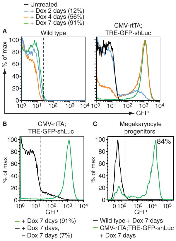
Reversible green fluorescent protein (GFP) reporter expression in the megakaryocyte/platelet lineage. (A) Flow cytometry analysis of GFP expression in CD41+ platelets in peripheral blood of representative wild type (left panel) and CMV-rtTA; TRE-GFP-shLuc bi-transgenic (right panel) mice after doxycycline treatment of indicated durations. Percent GFP+ cells in bi-transgenic samples are indicated. (B) Flow cytometry analysis of GFP expression in peripheral blood platelets of representative CMV-rtTA; TRE-GFP-shLuc mice after 1 week on doxycycline (green) followed by a week off doxycycline (black), showing reversible GFP expression in bi-transgenic platelets. PercentGFP+cells are indicated. (C) Flow cytometry analysis of doxycycline-induced GFP expression in Lin– Sca1–cKit+CD150+CD41+ megakaryocyte precursors isolated from bone marrow of wild type (black) and CMV-rtTA; TRE-GFP-shLuc bi-transgenic (green) mice. Percent GFP+ cells from bi-transgenic mice is indicated.

