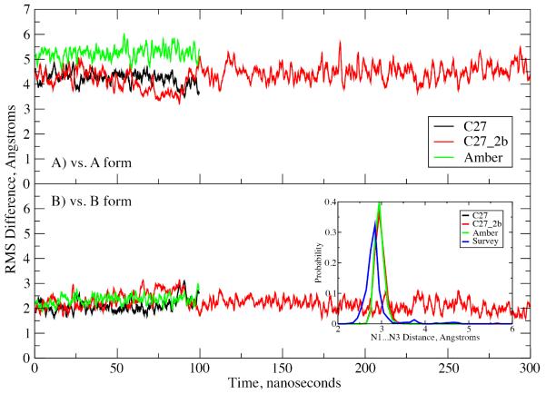
Figure 3.
RMS difference versus time for the EcoR1 dodecamer in solution. RMS differences vs. the A) canonical A form and B) canonical B form of DNA for all non-hydrogen atoms in the non-terminal residues. Results are for the C27 (black), C27_2b (red) and AMBER Parm99bsc0 (green) force fields. Inset: Watson-Crick base pair interaction based on the N1…N3 distance distributions for the three force fields and data from the survey of B form DNA crystal structures (blue).

