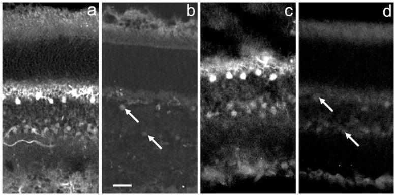
Fig. 2.
Comparison of anti-DARPP-32 IR in WT and DARPP-32-KO mouse retinas. (a and c) DARPP-32 IR in WT mouse retinas exposed, respectively, to (a) a mouse monoclonal and (c) a rabbit polyclonal anti-DARPP-32 antibody at 1 : 1000 dilution. (b and d) Immunostaining of the same antibodies in mouse knockout retinas. Retinal layers can be identified by reference to the labels in Fig. 1b. Note that in (c), the plane of the section is oblique. In the WT retinas, horizontal cells and amacrine cells are clearly labelled. In the KO retinas, these two cell types are faintly stained, as indicated by the arrows. Scale bar, 20 μm (applies to all panels).

