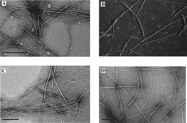
FIGURE 1.
Electron microscope images of hIAPP fibrils showing twisted morphology. A, negatively stained wild-type hIAPP fibrils. B, unilateral metal shadowing of wild-type hIAPP fibrils showing a left-handed twist (metal was deposited from the right; deposited metal is shown in white, and shadows are black). C, negatively stained singly labeled (13R1) hIAPP fibrils. D, negatively stained double-labeled (13R1/18R1) hIAPP fibrils. Bar: 200 nm.

