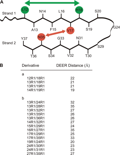
FIGURE 3.
Schematic model of hIAPP in protofilament and four-pulse DEER distance measurements. A, hIAPP with two β-strands regions connected by a loop. Placement of “inward” and “outward” oriented side chains is based on mobility measurements by continuous wave EPR (supplemental Fig. S3). Intrastrand (green) and interstrand (red) distances are indicated schematically. B, intramolecular DEER distances measured in doubly labeled hIAPP peptides within fibrils. Distances are from Gaussian fits of dipolar evolution data (supplemental Fig. S4).

