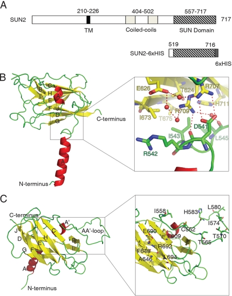
FIGURE 1.
Structure of the SUN domain monomer. A, schematic representation of the SUN2 C-terminal fragment that was used for the structure study. TM, transmembrane region. B and C, views of SUN domain monomer from two different directions. The secondary structural elements are labeled alphabetically from N terminus to C terminus. The right panel in B is the zoomed in image of the juncture area connecting the β-sandwich to the α-helix. The red balls represent water molecules involved in a hydrogen network (dashed lines, same below). The right panel in C shows a potential protein binding site located on the molecular surface of the β-sheet CHEF and the extended AA′ loop.

