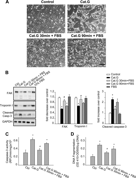
FIGURE 6.
Cat.G-induced myocyte anoikis is temporally associated with FA protein degradation. A–D, NRCMs were untreated, treated with Cat.G for 4 h, treated with Cat.G for 30 min followed by a washout, treated with Cat.G for 90 min followed by a washout, or treated with 10% FBS. A, phase contrast photomicrographs (Bar, 100 μm). B, representative immunoblot of FAK, troponin I and cleaved caspase-3. GAPDH was used as a loading control. Right: quantification of experiments expressed as mean ± S.E. from three separate cultures. *, p < 0.05 versus control, #, p < 0.05 versus Cat.G-treated cells. C and D, NRCMs were treated as indicated and assayed for caspase-3 activity using specific fluorogenic substrate or DNA fragmentation assay using anti-histone antibody ELISA. Results are expressed as relative fluorescence unit (RFU)/min/mg of proteins (C) or as (A410-A500)/mg DNA (D) for triplicate determinations from a single experiment (mean ± S.E.). *, p < 0.05 versus untreated control, #, p < 0.05 versus Cat.G-treated cells.

