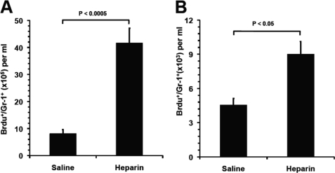
FIGURE 4.
Heparin released neutrophils from the bone marrow and demarginated neutrophils in the vasculature. A, neutrophil release from BM into blood circulation. BrdU-labeled BM neutrophils in blood as Gr-1+/BrdU+ cell were quantified 90 min post-heparin injection. B, heparin mobilized neutrophils from marginating pool to circulating pool in the vasculature. Transfused BrdU-labeled neutrophils (BrdU+/Gr-1+) were allowed to reach the dynamic equilibrium of distribution between circulating and marginating pools, and then the BrdU+/Gr-1+ cells in circulating pool were quantified 90 min post-heparin injection. The data were summarized from two sets of experiments with 5–6 mice per group. Each bar represents the average value ± S.E. The statistical analysis was carried by paired Student's t test in comparison with saline treatment.

