FIGURE 6.
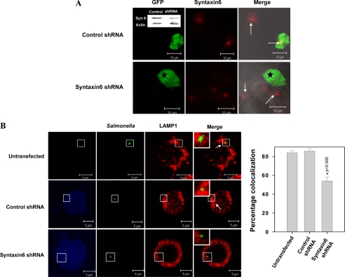
Recruitment of LAMP1 on SCP in Syntaxin6-depleted macrophages. A, confocal micrographs showing depletion of Syntaxin6 in Syntaxin6 shRNA-transfected cells. Luciferase shRNA-transfected cells were used as control. GFP expression indicates transfected cells (star) and Syntaxin6 appears in red (indicated by arrows). Inset, Western blot showing levels of Syntaxin6 in indicated cells. B, RAW 264.7 macrophages were transfected with respective shRNA and infected with WT:Salmonella as described under “Experimental Procedures.” The confocal image represents a single x/y plane of a Z-section where transfected cells are represented in blue; green shows the Salmonella and LAMP1 appears in red. Arrow indicates colocalization. Inset shows the enlarged region. The graph represents the percentage of SCP positive for LAMP1 from at least 50 observations. The level of significance was calculated by a t test, and the p value is as indicated.

