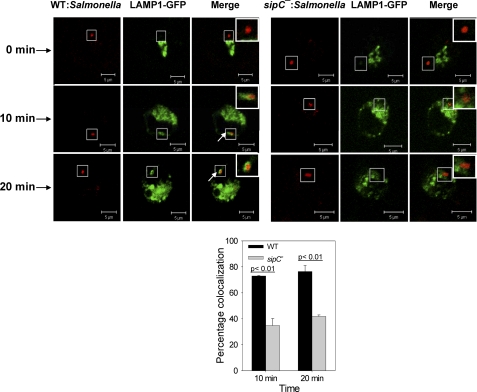
FIGURE 7.
Recruitment of LAMP1 on SCP by fusion with Golgi-derived LAMP1-containing vesicles. The image represents a single x/y plane of a Z-section, where green shows the LAMP1 and Salmonella appears in red. A ring-like staining around Salmonella indicates LAMP1-positive phagosomes as indicated by arrows. The graph below represents the percentage of SCP positive for LAMP1 where at least 100 phagosomes were analyzed for each time point. Data from three independent experiments was analyzed by t test, and levels of significance are indicated by p values. Inset shows the enlarged region.

