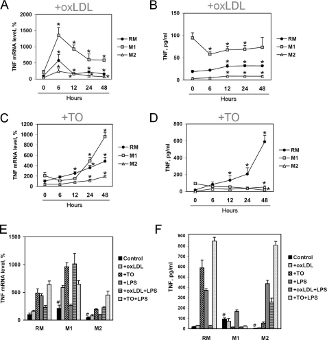
FIGURE 5.
TNFα expression and secretion by human resting and classically (M1) and alternatively (M2) activated PBM-derived macrophages. A and B, level of TNFα mRNA (A) determined by real-time RT-PCR and TNFα secretion (B) determined by ELISA in resting and classically or alternatively activated PBM-derived macrophages 0–48 h after the addition of oxLDL (50 μg/ml). C and D, the level of TNFα mRNA (C) determined by real-time RT-PCR and TNFα secretion (D) determined by ELISA in resting and classically or alternatively activated PBM-derived macrophages 0–48 h after the addition of LXR agonist TO191317 (TO; 2.5 μm). E, level of TNFα mRNA in human PBM-derived macrophages; real-time RT-PCR; 100% is the level in the macrophages treated with BSA. F, ELISA of TNFα protein content in the culture medium of PBM-derived macrophages. PBM-derived macrophages were treated with BSA (20 ng/ml) (RM), with IFNγ (20 ng/ml) and bacterial LPS (100 ng/ml) (M1) or IL-4 (20 ng/ml) (M2) for 6 days. Cells were treated with oxLDL (50 μg/ml) and/or LXR agonist TO191317 (2.5 μm) and/or LPS (100 ng/ml) for 48 h. Values are presented as means ± S.E. (error bars) of six independent experiments. The statistical analyses of differences were performed separately for values in the RM, M1, or M2 group using an unpaired Student's t test (*, p < 0.05) or Dunnett's criterion (for comparison of RM, M1, and M2) (#, p < 0.05).

