FIGURE 4.
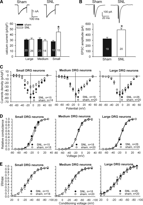
Effects of nerve injury on HVACC activity of DRG neurons and monosynaptic EPSCs of spinal dorsal horn neurons. A, original current traces (upper panel) show HVACC currents in small DRG neurons from a control and a nerve-injured rat. Group data (lower panel) show that SNL increased HVACC currents in small, but not large and medium, DRG neurons. Neurons were voltage-clamped at −90 mV and depolarized to 0 mV for 200 ms. The number of cells in each group is indicated in the column. B, representative traces and group data show the monosynaptic EPSCs of lamina II neurons in spinal cords obtained from sham control (n = 19) and SNL (n = 20) rats. C, effect of SNL on the current-voltage relationship of HVACCs in small, medium, and large DRG neurons. D, effect of SNL on voltage-dependent activation of HVACCs in small, medium, and large DRG neurons. Note that SNL shifted voltage-dependent activation of HVACCs to the left only in small DRG neurons. The V0.5 in control and SNL rats was −13.7 ± 0.1 and −17.5 ± 0.1 mV (p < 0.05), respectively. The slope factor in control and SNL rats was 8.33 ± 0.2 and 7.80 ± 0.1 mV (p > 0.05), respectively. E, effect of SNL on voltage-dependent inactivation of HVACCs in small, medium, and large DRG neurons. *, p < 0.05, compared with the corresponding value in the sham group. pF, picofarad.

