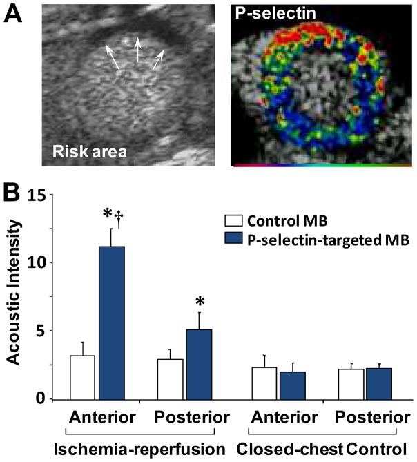
Figure 4.
Contrast echocardiographic imaging of recent myocardial ischemia with P-selectin targeted microbubbles.(21) (A) Short axis contrast echocardiographic images from a mouse illustrating hypoperfusion (arrows) of the anterior wall on perfusion imaging during brief occlusion of the LAD (left) and enhancement within the previously ischemic anterior territory with P-selectin-targeted microbubbles injected intravenously 45 minutes after reflow. Color scale at bottom. (B) Qauntitative data of signal enhancement from P-selectin microbubbles in the LAD (anterior) territory in control mice and in mice 45 min after ischemia-reperfusion injury.

