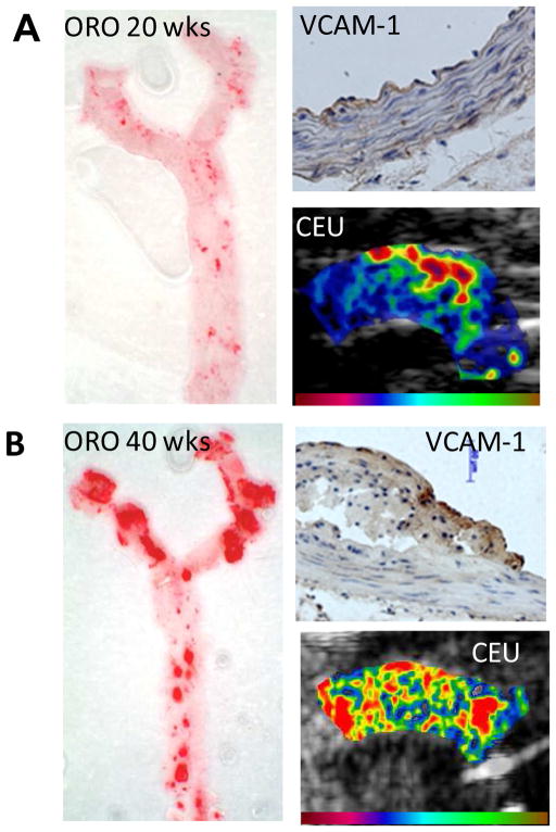
Figure 5.
Molecular imaging of VCAM-1 to detect endothelial activation in early atherosclerotic disease.(25) Images are from a model of age-dependent atherosclerosis in mice (A) at 10 wks an (B) at 40 weeks of age. En face images of the thoracic aorta with Oil Red-O (ORO) staining demonstrates age-related increase in spatial extent of plaque formation. Immunohistochemistry shows VCAM-1 expression despite only mild intimal thickening at 10 wks whereas there is dense VCAM-1 expression and large lesion formation at 40 wks. Contrast-enhanced ultrasound (CEU) molecular imaging of VCAM-1 shows enhancement in the aortic arch at both stages, although signal at the early time point was mostly localized to the region where future plaque formation is most severe which is at the origin of the brachiocephalic artery.

