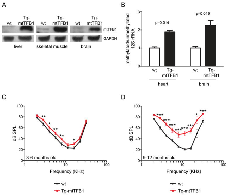
Figure 4. Transgenic mice that over-express the mtTFB1 rRNA methyltransferase exhibit 12S hyper-methylation and progressive hearing loss.
(A) Western blot of mtTFB1 in wild-type (wt) and transgenic mtTFB1 (Tg-mtTFB1) mouse tissues using GAPDH as loading control.
(B) Quantification of the degree of 12S rRNA methylation in Tg-mtTFB1 mouse heart and brain tissues using a methylation-sensitive mitochondrial 12S rRNA primer-extension assay (Cotney et al., 2009). The values plotted represent mean of methylated/unmethylated ± standard deviation (n=3), with t-test p-values indicated.
(C) Hearing thresholds determined by ABR analysis of Tg-mtTFB1 (red points) and control wild-type (wt), non-transgenic littermate control mice (black points) are shown in the audiogram (frequency vs. threshold curve). The threshold (in sound pressure level, dB SPL) was tested with 5-dB resolution and frequencies in half octave steps from 32 to 2 kHz. The values plotted represent mean ± standard error of the mean, with t-test p-values indicated (*, p<0.05; **, p<0.01; ***, p<0.001). Mice were tested at ages 3–6 months (9 wt mice and 12 Tg-mtTFB1 mice were used).
(D) same as in (C) except mice were tested at ages 9–12 months (5 wild-type mice and 6 Tg-mtTFB1 mice were used).

