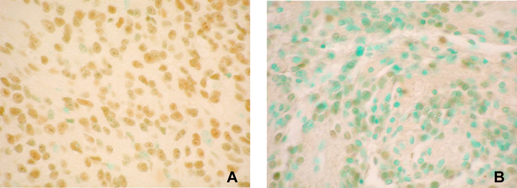
Figure 2. Ape1/Ref-1 immunoreactivity in grade II ependymoma cells.
Tumor nuclei expressing detectable Ape1/Ref-1 are brown (stained with 3,3’-diaminobenzidine tetrachloride); negative nuclei are green (stained with methyl green). Fraction of immmunopositive cells was 82% for A and 40% for B. All images 40X power.

