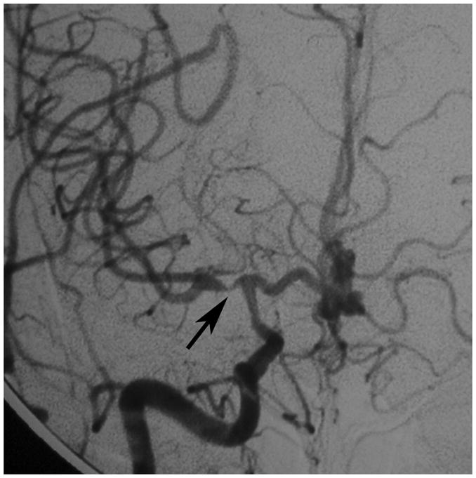
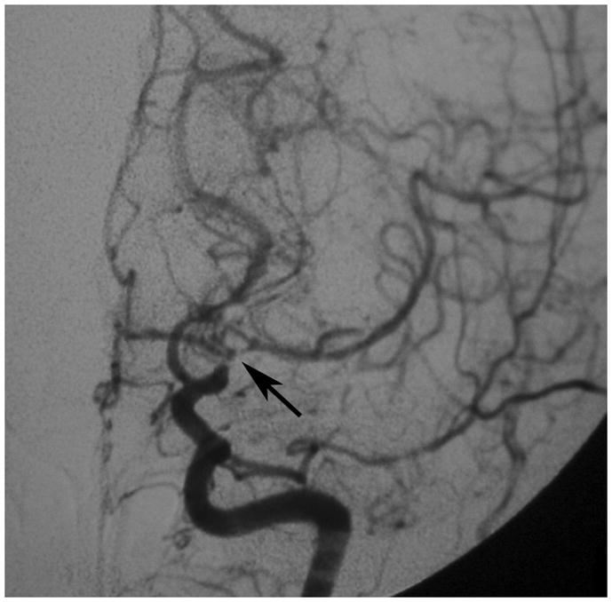
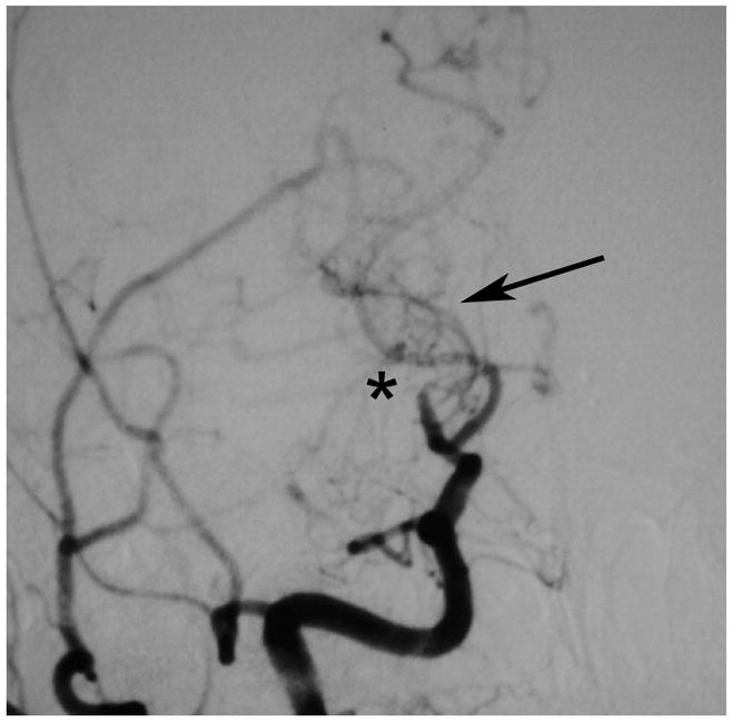
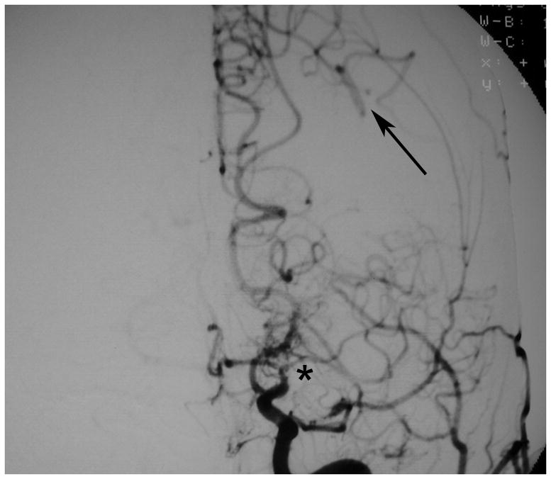
Figure 1. Development of moyamoya collaterals in a North American adult.


Legend: A 45 year old female patient initially presented with ischemic stroke. Cerebral angiography demonstrated proximal stenosis of the middle cerebral arteries bilaterally (A and V). Figure 1A, AP projection after right common carotid artery injection, demonstrates severe narrowing of the proximal middle cerebral artery (black arrow). Figure 1B, AP projection after left common carotid artery injection, shows similar changes of the distal internal and proximal middle and anterior cerebral arteries (black arrow). She was treated with steroids for presumed vasculitis. On repeat angiography approximately 6 months later, the stenotic lesions markedly progressed with the interval development of moyamoya collaterals (C and D). Figure 1C, AP projection after right common carotid artery injection, shows occlusion of the distal internal carotid artery (asterisk) with moyamoya collaterals (black arrow). Figure 1D, AP projection after left common carotid artery injection, demonstrates similar changes (middle cerebral artery occlusion with moyamoya collateral formation, asterisk) with pial collaterals from the posterior artery branches to the middle cerebral artery territory (black arrow).
