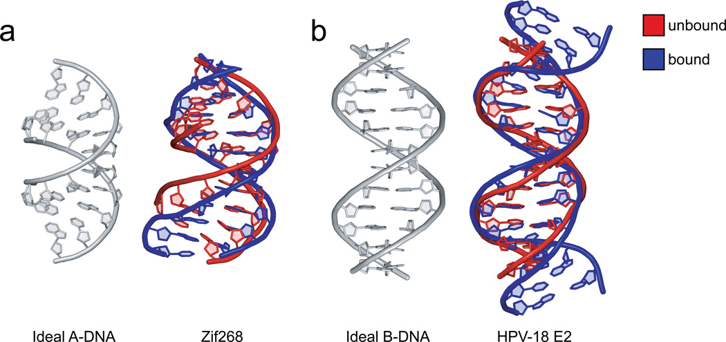
Figure 3. DNAs bound to proteins have features already present in unbound DNAs.
a. The structure of the unbound FIN-B sequence (PDB ID 2b1c) is similar to ideal A-DNA (grey), while the bound structure of the Zif286-DNA complex (PDB ID 1a1f) has some A-DNA characteristics, notably a wider minor groove than normally found in B-DNA.
b. The specific HPV-18 E2 site (PDB ID 1ilc) contains an A-tract AATT in the central region of the helix, which, although not contacted by the protein, bends the free-DNA structure (red) in a similar manner as seen in the bound structure (blue) of the HPV-18 E2-DNA complex (PDB ID 1jj4). In comparison to ideal B-DNA (grey), the bending is reflected by a minor groove narrowing in the center of the free and bound DNA.

