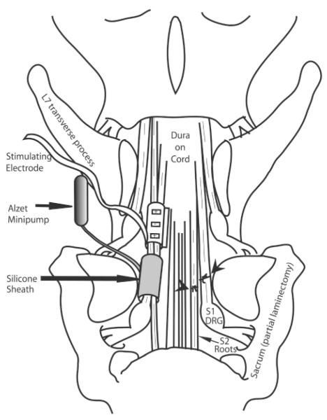
FIG. 1.
Diagram of the surgical repair methodology. A spinal laminectomy was performed, removing lumbar 6 (L6), and part of L7 and sacrum 1 (S1) lamina and spinous processes. The site of ventral and dorsal root transection and surgical repair is shown on the right side of the diagram (indicated by arrowheads). The placement of the stimulating electrode sheath and silicon sheath receiving brain-derived neurotrophic factor (BDNF) from the Alzet minipump is shown on the left side of the diagram. DRG, dorsal root ganglion.

