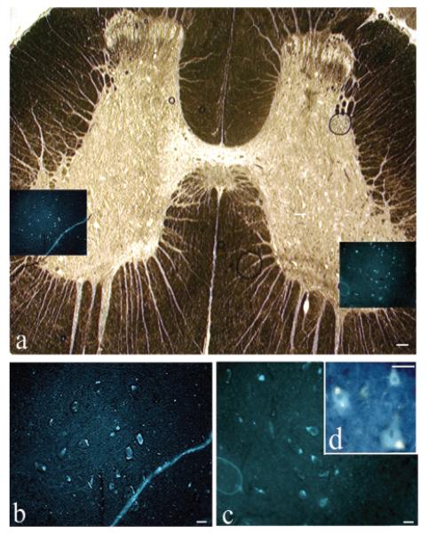
FIG. 3.
Reinnervation of the bladder from the cord. Fluorogold retrogradely labeled neuronal cell bodies in spinal cord following injection into the bladder 3 weeks before euthanasia, which was 12 months postoperative. (A) Cross-section of a sacral cord segment. A bright-field low-magnification photomicrograph of an unstained (other than the fluorogold, which is not visible in bright field) wet-mounted cord that had been cover slipped with 80% glycerol in phosphate-buffered saline (PBS). Insets in A show fluorogold (FG)–labeled neuronal cell bodies located bilaterally in lateral ventral horns. (B,C) Enlargement of insets in panel A showing fluorogold-labeled neuronal cell bodies. (D) Higher power of neuronal cell bodies showing cytosolic localization of fluorogold dye in neuronal cell bodies. Scale bar 100 μm (A), 50 μm (B,C).

