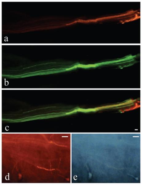
FIG. 4.
Postmortem labeling of ventral and dorsal root axons. Photomicrographs show a root immediately proximal to the repair site. (A) Postmortem retrograde labeling of axons (red DiI) is visible in the ventral root, as determined anatomically, after dye injection into the spinal nerve 2 cm distal to the ventral root transection/repair site. (B) Postmortem anterograde labeling of axons from the spinal cord (green DiO) is visible in the same ventral root as shown in A. (C) A merger of both A and B, and shows overlap of these 2 dyes. (D,E) High-power pictures from sacral spinal cord segments showing two fluorogold-labeled motor neuronal cell bodies that were retrogradely labeled from the bladder (arrows in D). These same neurons are also labeled with DiI which had been inserted into a ventral root distal to the site of reanastomosis (E). Scale bar = 50 μm.

