
FIG. 7.
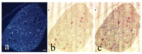
Number of new axons in repaired root. Fluorogold (FG)–labeled axons in ventral and dorsal roots. (A–C) Cross-section of a root as it nears the cord, a root region that is proximal to the site of reanastomosis. (A) Small fluorescence blue profiles in root are fluorogold (FG) retrogradely labeled axons (labeled after injection into the bladder). (B) Bright-field image of same root; red filled circles are blood vessels. (C) Images in panels A and B are shown superimposed. Scale bar = 50 μm.