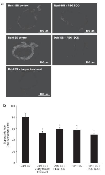
Figure 1.
Superoxide levels in SS and Ren-1 BN rats. (a) Representative fluorescent images of basilar artery cross-sections (10 μm thickness) from Dahl salt-sensitive (SS) and Ren1-Brown Norway (BN) rats ± 15 min preincubation with 100 U/ml polyethylene glycol (PEG) superoxide dismutase (SOD) in the presence of 5 μmol/l dihydroethidium (DHE). a separate group of Dahl SS rats was treated with tempol (15 mg/kg/day) for 7 days. Images were taken at ×20 magnification. Bar = 100 μm. (b) Vascular superoxide levels quantified as fluorescence units in DHE-treated basilar artery cross-sections from Dahl SS (n = 5) and Ren1-BN (n = 6) rats ± 100 U/ml PEG SOD (n = 6, 5 respectively). a separate group of Dahl SS rats was treated with tempol for 7 days (n = 8). Data are expressed as mean value (raw fluorescence units) ± s.e.m. *Significant difference (P < 0.05) all groups vs. Dahl SS.

