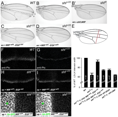
Figure 7. Vertebrate WIF domain regulates long-range Hh signaling.
(A) Wild type wing showing the positions of the first through fifth longitudinal veins (L1–L5) and the position of the A/P compartment boundary (dashed line). (B) Reduced L3–L4 spacing in shf mutant wing (B) is not improved upon expression of UAS-shfΔWIF (B′). (C, D) Posterior, en-Gal4-driven expression of UAS-WIFwif1-EGFshf (C) or UAS-WIFshf-EGFwif1 (D) in shfx33/Y wings. UAS-WIFwif1-EGFshf strongly improved and UAS-WIFshf-EGFwif1 weakly improved L3–L4 spacing. (E) Comparison of L3–L4 spacing in wild type, shf2, and shfx33/Y with en-Gal4-driven UAS-construct expression. To compensate for differences in overall wing size, we normalized the L3–L4 distance to the distance between the anterior and posterior margins (red bars). In all but one case we presented the experimental normalized L3–L4 distances as percentages of the wild type normalized distances. However, since expression of UAS-WIFwif1-EGFshf reduced the size of the posterior compartment, and thus the distance between the anterior and posterior margins, we compared the normalized L3–L4 distance in shfx33 UAS-WIFwif1-EGFshf wings to the normalized L3–L4 distance in non-shf UAS-WIFwif1-EGFshf siblings (n = 31). Bars denote standard deviation. Two-tailed Student's t test showed no significant differences between UAS-WIFshf-EGFwif1 and UAS-shfΔEGF, or between shf2 and shf2, UAS-shfΔWIF. Differences between the other conditions were significant (p<0.0001). (F–I) Anti-Ptc staining in wild type (F), shfx33/Y (G), and shfx33/Y wing discs with ap-Gal4-driven expression of UAS-WIFwif1-EGFshf (H) or UAS-WIFshf-EGFwif1 (I). The improvement in the width of Ptc expression in shfx33 discs was stronger after expression of UAS-WIFwif1-EGFshf than UAS-WIFshf-EGFwif1. (J, K) shfx33/Y wing discs with ap-Gal4-driven expression of UAS-hh-GFP and UAS-WIFwif1-EGFshf (J) or UAS-hh-GFP and UAS-WIFshf-EGFwif1 (K). UAS-WIFwif1-EGFshf strongly improved the ventral accumulation of Hh-GFP, while the improvement with UAS-WIFshf-EGFwif1 was more modest (quantifications in Figure S9).

