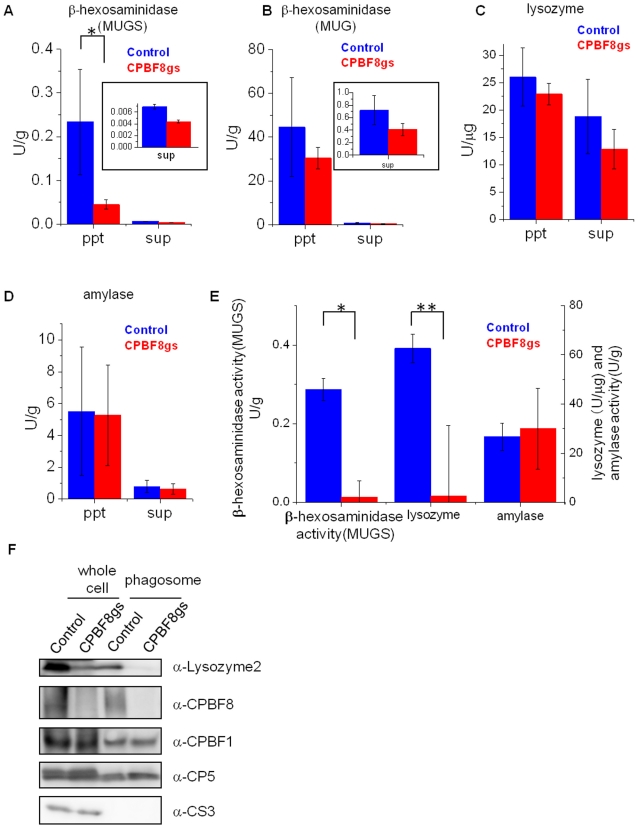
Figure 4. Enzymatic activities in total lysates, culture supernatant, and phagosomes and immuno blot analysis of total lysates and phagosomes derived from control and CPBF8gs strains.
After the amoebas were incubated in fresh medium for 2 h, trophozoites and culture supernatant were separated by brief centrifugation. After the supernatant was removed (“sup”), the cell pellet was resuspended and solublized in lysis buffer (“ppt”). To isolate phagosomes (E), the amoebas were incubated with latex beads, and, after brief centrifugation, the cell pellet was resuspended, and mechanically homogenized. The phagosomes were isolated by ultracentrifugation on a sucrose gradient. Enzymatic activities of culture supernatant, whole lysate (A–D), and phagosomes (E) are shown. (A–D) Enzymatic activities of β-hexosaminidase activity toward MUGS (A) and MUG (B), lysozyme activity (C), and amylase activity (D) in the cell pellet and culture supernatant. (E) Enzymatic activities of β-hexosaminidase toward MUGS, lysozyme, and amylase in phagosomes. Data shown are the means ± standard deviations of three independent experiments. “*” or “**” represents statistical significance at p<0.01 or p<0.05, respectively. (F) Immuno blot analysis of the cell pellet and the phagosome fraction. Approximately 20 µg of the cell pellet and 2 µg of the phagosome fraction were electrophoresed. CPBF1 and CP5 are phagosomal proteins, while cysteine synthase 3 (CS3) is a cytosolic marker. Abbreviations are: ppt, pellet fraction; sup, culture supernatant; phagosome, phagosome fraction.

