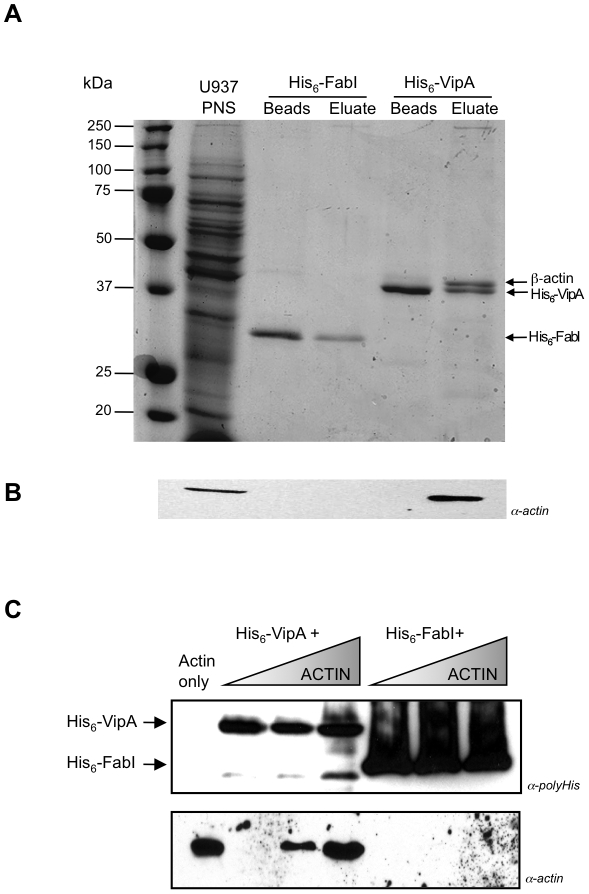
Figure 1. In vitro interaction analysis between His6-VipA and actin by pull-down assays.
A. U937 cell Post-Nuclear Supernatants (U937 PNS) were incubated with Ni-NTA agarose beads preloaded with His6-VipA or His6-FabI (see Materials and Methods for details). After washing, bound proteins were eluted with 500 mM Imidazole. Samples containing preloaded beads or eluates were resolved by SDS-PAGE and bands visualized by Coomassie staining. The differential band appearing in the VipA eluate (arrow) was excised and identified by LC/MS as β-actin. B. Similarly prepared samples were resolved by SDS-PAGE, transferred to nitrocellulose and immunoblotted with an anti-actin monoclonal antibody (α-actin). C. Ni-NTA agarose beads preloaded with His6-FabI or His6-VipA were incubated with increasing concentrations of purified G-actin, washed and eluted. Eluates were resolved by SDS-PAGE, transferred to nitrocellulose and Western-blotted with an anti-histidine antibody (α-polyHis, panel above) or an anti-actin monoclonal antibody (α-actin, panel below).

