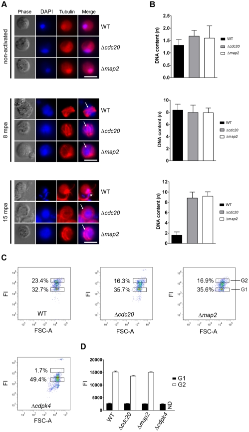
Figure 4. Analysis of genome replication in activated male gametocytes by direct (immuno) fluorescence microscopy and FACS analysis.
A. Direct immunolabelling of α-tubulin (red) and DNA (Hoechst – blue) in activated gametocytes fixed at different time points post activation (pa). Representative cells from one of three experiments are shown. The 8 min time point shows characteristic axonemic circling of the nucleus (arrow) whereby each of the 8 axonemes lie completely within the cytoplasm, coiled around the nucleus. The 15 min time point illustrates an exflagellating wild-type microgametocyte with condensed DNA entering the flagella (indicated by *). Δcdc20 and Δmap2 parasites are arrested at the exflagellation stage. Bar = 5 µm. mpa = minutes post-activation. B. Fluorometric anaylsis of DNA content (n) after DAPI nuclear staining. Microgametocytes were at 0 mpa (non-activated), 8 mpa or 15 mpa. The mean DNA content (and standard deviation) of 10 nuclei per sample are shown. Values are expressed relative to the average fluorescence intensity of 10 haploid ring-stage parasites from the same slide [69]. All values were corrected for background fluorescence. C. Determination of DNA content of purified, activated male gametocytes at 8 mpa by FACS analyses of Hoechst-stained parasites [63]. Dot plots show the mean percentage of gametocytes in gate G1 (inactivated and activated female gametocytes) and G2 (activated gametocytes with an 8n DNA content. Wild-type, Δcdc20 and Δmap2 parasites show a high percentage of activated male gametocytes with an 8n DNA content (see D). The previously characterised Δcdpk4 parasites [28] were used as a control as they do not undergo DNA replication upon activation. FI = Fluorescence Intensity. D. Mean Hoechst fluorescence intensity (DNA content) (±SD) of gametocytes in gates G1 and G2 in three independent experiments. The DNA content of Δcdc20 and Δmap2 male gametocytes (gate G2) at 8 mpa is comparable to that of wild-type gametocytes whereas activated, DNA replicating males are absent in Δcdpk4 parasites. FI = Fluorescence Intensity; ND = not determined.

