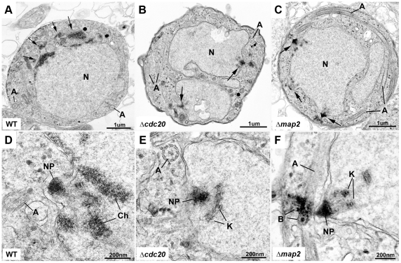
Figure 5. Ultrastructural analysis of Δcdc20 and Δmap2 parasite lines.
Electron micrographs of wild-type (A, D), Δcdc20 (B, E) and Δmap2 (C, F) gametocytes 15 min after activation. A. Late stage wild-type microgametocyte exhibiting areas of electron dense chromatin (arrows) budding from the nucleus (N). A = axonemes. B and C. Mutant microgametocytes at an early stage in microgametocyte development showing multiple nuclear poles (arrows) with radiating microtubules. N = nucleus; A = axoneme. D. Detail from A. showing the nuclear pole (NP) and areas of electron dense chromatin (Ch). A = axoneme. E and F. detail from the two mutants (B, C) showing the nuclear pole (NP) with radiating microtubules and attached kinetochores (K) but an absence of condensed chromatin. A = axoneme; B = basal body.

