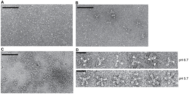
Figure 6. Electron microscopy on negatively stained Gth at pH 7.5, pH 6.7 and pH 5.7.
(A) At pH 7.5, no oligomer of Gth is detected and a definite molecular shape cannot be identified. (B) At pH 6.7 a few Gth assemblies can be detected. (C) At pH 5.7 Gth trimers are observed assembled either in lattice via lateral interactions or in rosette-like assemblies. Protein concentration was 0.1 mg/mL for all 3 samples. The scale bar is for 100 nm. (D) Close up view of Gth rosettes observed at pH 6.7 and 5.7. Notice the differences in the aspect of the protein assemblies. The scale bar is for 20 nm.

