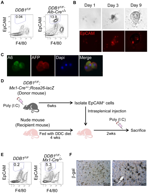
Figure 4. EpCAM-positive cells from DDB1 mutant liver proliferate in vitro and repopulate the liver in vivo.
(A) Analysis by flow cytometry of EpCAM+ F4/80− cells in non-parenchymal fractions prepared from 4-week old DDB1F/F and DDB1F/F;Alb-Cre+/+ mice. The ratio of EpCAM+ F4/80− cells from a representative experiment is shown in each panel. (B) In vitro culture of EpCAM+ cells sorted by FACS from DDB1 mutant liver and seeded on Matrigel. Cells form colonies after 1, 3, and 9 days of culture (upper panels) and exhibit EpCAM positivity (lower panels). (C) Co-IF staining for A6 and AFP on colonies from cultured EpCAM+ cells. (D) Experimental scheme for in vivo differentiation of EpCAM+ OCs. Adult DDB1F/F;Mx1-Cre+/−;Rosa26-lacZ mice were generated and injected with poly(I:C). EpCAM+ cells were isolated by FACS 6 weeks later, and injected intrasplenically to nude mice that had been fed with DDC diet. The recipient nude mice received poly(I:C) injection 2 weeks later and liver collected for analysis on the following day. (E) Isolation of EpCAM+ F4/80− cells from donor or control mouse liver. The ratio of EpCAM+ F4/80− cells is shown in each panel. (F) IHC staining for ß-gal on liver sections from recipient nude mice. Arrows indicate positively stained hepatocytes.

