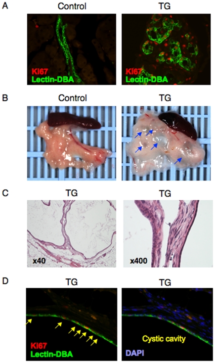
Figure 3. Enhanced proliferation of pancreatic duct epithelial cells and development of polycystic pancreas in TG mice.
(A) Immunohistochemistry with anti-Ki67 antibody (red) along with Lectin-DBA (green) was conducted in pancreatic sections from 2 month old TG and control mice. Representative results are shown. (B) Photographs of the pancreas in 12 month old TG and control mice. Representative pictures are shown. (C) Hematoxylin and eosin (HE) staining (left panel indicates ×40 magnification and right ×400) in pancreatic sections from 12 month old TG mice. Representative images are shown. (D) Immunohistochemistry with anti-Ki67 antibody (red) along with Lectin-DBA (green) was conducted in pancreatic sections from 12 month old TG mice. Representative images of pancreatic cysts are shown. Arrow indicates Ki67 positive cells.

