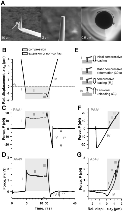
Figure 1. Illustration of the 4-step protocol based on FE-AFM tips used to probe cell mechanoresponses to compression and extension.
(A) Representative SEM images of a nanofabricated cylindrical FE-AFM tip. A whole FIB-milled cylindrical tip is shown in the left panel, and detailed lateral and top view images of the tip are shown in the middle and right panels, respectively. (B) Driving signal of the piezotranslator in z as a function of time (t) used to probe the sample mechanoresponse to compression and extension. Note that the z axis was scaled relative to zc, which is marked with an horizontal dashed line. Although the relative z signal started at 13.5 µm, the z axis was limited to 4 µm above zc to better visualize the range corresponding to step III and IV. A break in the t axis was introduced for the same purpose. Corresponding F recordings as a function of t on a PAA+ gel and a single A549 cell are shown in (C) and (D), respectively. A common t axis was used in (B–D). F* was obtained from step IV as illustrated in (C, D). (E) Cartoon describing the tip-sample mechanical interactions corresponding to the 4-steps of the experimental protocol. EC and ET were calculated using signals from step III and IV. F signals from (C) and (D) were plotted against z in (F) and (G), respectively. The parts of the z and F signals obtained in compression were highlighted in gray. All F data were scaled relative to the corresponding zero force (k⋅d0).

