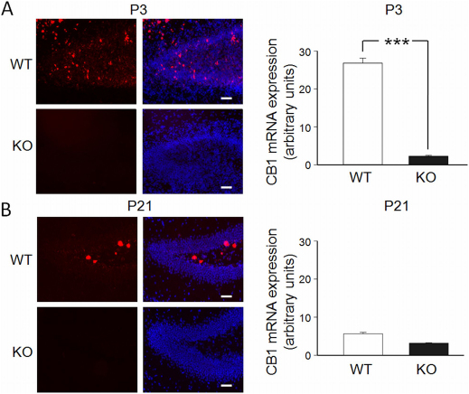
Figure 8. Developmentally-regulated expression of CB1 mRNA in the granule cell layer of the dentate gyrus.
(A) and (B) On the left, photomicrographs representing CB1 mRNA staining (red) alone or together with DAPI nuclear counterstaining (blue) in the dentate gyrus of WT and CB1-KO mice at P3 (A) and at P21 (B) animals. Cal. bar: 20 μm. On the right, Each column represents the averaged value of CB1 mRNA expression obtained in 4-8 sections corresponding to dorsal DG of WT or CB1-KO mice at P3 and at P21 (2 mice per group; see methods). ***p<0.001.

