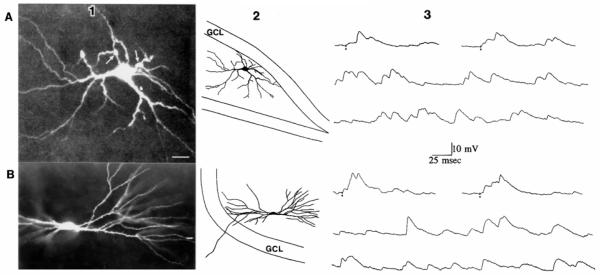
FIG. 1.
Spontaneous and evoked excitatory postsynaptic potentials (EPSPs) of different spiny hilar cells are similar. A: EPSPs of a spiny hilar “mossy” cell. A1: spiny hilar cell with excrescences on its cell body and proximal dendrites is shown. This image is a composite of 15 optical sections taken with a confocal microscope through a slice. Calibration = 15 μm. A2: drawing of the cell shown in A1 illustrates where the cell was located in the slice. Dorsal is up, area CA3 is to the left. The granule cell layer (GCL) is outlined. A3: evoked (top) and spontaneous (middle and bottom) EPSPs of the cell shown in A, 1 and 2. Small circles: stimuli to the outer molecular layer. Membrane potential = −64 mV. B: EPSPs of a spiny hilar cell without excrescences. Bl: spiny hilar cell with simple spines is shown. The cell was in a different slice from the cell in A. Calibration is the same as in A. B2: drawing of the entire cell and its location in the hilus. Dorsal is down, area CA3 is to the right. A portion of the upper blade of the dentate gyrus is outlined. B3: evoked (top) and spontaneous (middle and bottom) EPSPs of the cell shown in B.1 and 2. Stimuli were applied to the outer molecular layer. Membrane potential = −63 mV.

