FIG. 5.
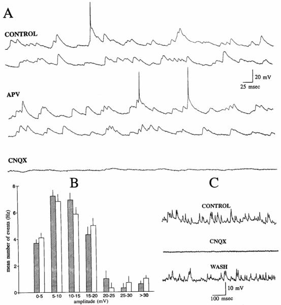
Blockade of spontaneous activity of a spiny hilar cell by the α-amino-3-hydroxy-5-methyl-4-isoxazolepropionic acid (AMPA)/kainate receptor 6-cyano-7-nitroquinoxaline-2,3-dione (CNQX) but not the N-methyl-d-aspartate NMDA) receptor antagonist 2-amino-5-phosphonovaleric acid (APV). A: CONTROL: continuous record of spontaneous activity of a spiny hilar cell. Membrane potential = −60 mV. APV: continuous record of spontaneous activity from the same cell as in A. recorded 45 min after 50 μM APV was added to the perfusate. Same membrane potential as in control. CNQX: continuous record from the same cell, recorded 15 min after 5 μM CNQX was added to the perfusate. CNQX was added at approximately the same time that the record in APV (shown above) was sampled. Same membrane potential as in control. B: histogram shows the mean number of spontaneous EPSPs (Hertz; mean ± SE) for the cell in A, in control (shaded bars), and 45 min after perfusion with APV (clear bars). C: spontaneous activity of a spiny hilar cell before 5 μM CNQX application (CONTROL), during bath application (CNQX), and 50 min after CNQX-free buffer was reintroduced to the slice (WASH).

