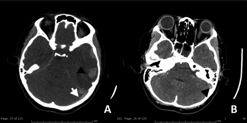
Figure 2.
(A) Axial computed-tomography noncontrast study) demonstrated hyperdensity of acute thrombus in left transverse sigmoid sinus (white arrow) with mixed hypo-hyperdensity lesion at left parieto-temporal region, measured about 4.9×2.8 cm in size (arrowhead). (B) Contrast enhanced computed-tomography scan shows filling defect in left transverse-sigmoid sinus (black arrow). All of these findings are suggestive of left sigmoid and transeverse sinovenous sinus thromboses with acute intracerebral hemorrhage.

