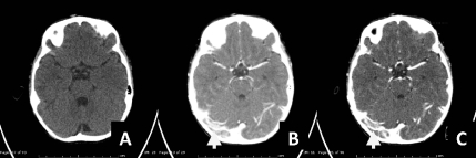
Figure 6.
Axial computed-tomography noncontrast study revealed A) isodensity lesion at right transverse sinus in non contrast image which showing as filling defect along right transverse sinus, right sigmoid sinus and right jugular bulb in post contrast image B,C), suggestive of partial venous sinus thrombosis.

