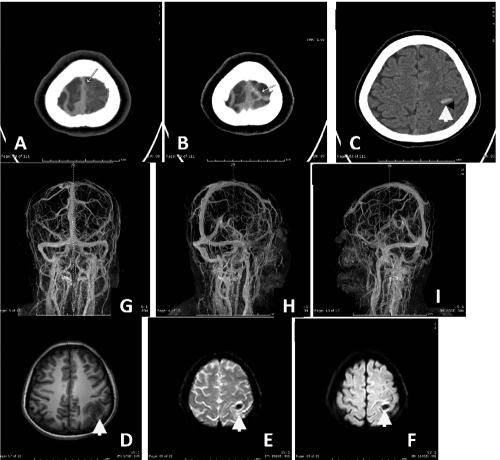
Figure 9.
Axial noncontrast computed-tomography scan revealed A,B,C) hyperdense of cord sign in left cortical vein with noncontrast filling in post contrast enhanced study. Hemorrhagic spot in left parietal lobe is noted (arrow). Magnetic resonance imaging shows D,E,F) hyposignal T1W, T2W_FFE (shows blooming susceptibility effct (arrow) of hemorrhage in left parietal lobe. Post contrast enhanced MR venography showed G,H,I) a small filling defect at superior sagittal sinus (seen on 3D_T1W/Gd) and lacking of cortical vein of left high parietal region corresponding with CT imaging, compatible with cerebral venous thrombosis.

