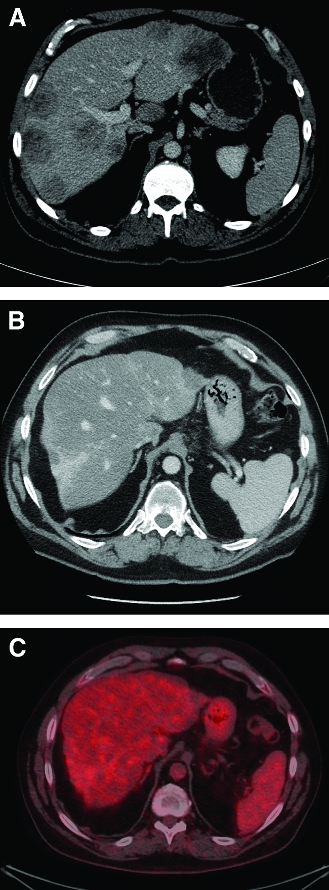
Figure 1.

Liver metastases before and after combination chemotherapy. (A): Pretreatment portal venous phase computed tomography (CT) scan showing extensive bilobar liver metastases. (B): Post-treatment portal venous phase CT scan showing fibrosteatotic replacement of metastases and nodular, contracted, steatotic liver, consistent with chemotherapy-associated steatohepatitis. Patient had received 12 months of capecitabine, irinotecan, and bevacizumab. (C): Fused CT–18F-fluorodeoxyglucose positron emission tomography scan after 12 months of chemotherapy showing complete response of liver metastases.
